
Follow Us
© 2026 All Rights Reserved
 Skip to content
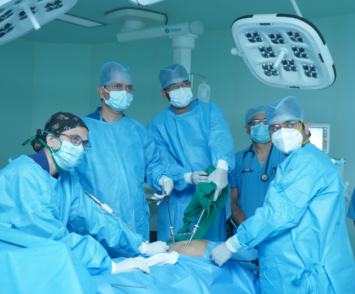
 Laparoscopy is a type of surgical procedure that allows a surgeon to access the inside of the abdomen (tummy) and pelvis without having to make large incisions in the skin. This procedure is known as keyhole surgery or minimally invasive surgery. Large incisions are avoided during laparoscopy because the surgeon uses an instrument called a laparoscope. This is a small rod like structure that has a light source and a camera, which relays images of the inside of the abdomen or pelvis to a television monitor.
The advantages of this technique over traditional open surgery are both to the surgeon and patient:
Laparoscopy can be used to help diagnose a wide range of conditions that develop inside the abdomen or pelvis. It can also be used to carry out surgical procedures, such as removing a damaged or diseased organ, or removing a tissue sample for further testing (biopsy).
Laparoscopy is most commonly used in:
Laparoscopy is carried out under general anaesthesia, so you won’t feel any pain during the procedure. During laparoscopy, the surgeon makes one or more small incisions in the abdomen. These allow the surgeon to insert the laparoscope, small surgical tools, and a tube used to pump gas(Co2) into the abdomen. This makes it easier for the surgeon to look around and operate.
After the procedure, the gas is let out of your abdomen, the incisions are closed using stitches and a dressing is applied.
You can often go home on the same day of your laparoscopy, although you may need to stay in hospital overnight.
Laparoscopy is a commonly performed procedure and serious complications are rare.
Minor complications are estimated to occur in one or two out of every 100 cases following laparoscopy.
They include:
Serious complications after laparoscopy are estimated to occur in one out of every 1,000 cases. They include:
Further surgery is often required to treat many of these more serious complications.
When it’s used
Laparoscopy is used to diagnose or treat numerous conditions.
During the procedure, small surgical instruments and devices are inserted through small incisions. This helps your surgeon perform whatever surgical procedure needs to be carried out.
Laparoscopy is used to diagnose or treat numerous conditions.
During the procedure, small surgical instruments and devices are inserted through small incisions. This helps your surgeon perform whatever surgical procedure needs to be carried out.
It’s often possible to diagnose a condition using detailed clinical examination and using non-invasive methods, such as an ultrasound scan, computerised tomography (CT) scan or magnetic resonance imaging (MRI) scan. Sometimes, however, the only way to confirm a diagnosis is to directly study the affected part of the body using a laparoscope.
Laparoscopies are now widely used to diagnose many different conditions and investigate certain symptoms. For example, they may be used for:
Laparoscopy can also be used to diagnose certain types of cancers. The laparoscope is used to obtain a sample of suspected cancerous tissue, so it can be sent to a laboratory for testing. This is known as a biopsy.
Cancers that can be diagnosed using laparoscopy include:
Laparoscopic surgery can be used to treat a number of different conditions, including:
Laparoscopy is performed under general anesthesia, so you’ll be unconscious during the procedure and have no memory of it. You can often go home on the same day.
Depending on the type of laparoscopic procedure being performed, you’ll usually be asked not to eat or drink anything for 6-12 hours beforehand.
If you’re taking blood-thinning medication (anticoagulants), such as aspirin or warfarin, you may be asked to stop taking it a few days beforehand. This is to prevent excessive bleeding during the operation. If you suffer from constipation, may be advised to take laxatives prescribed by your doctor the night before surgery.
If you smoke, you may be advised to stop during the lead-up to the operation. This is because smoking can delay healing after surgery and increase the risk of complications such as infection.
Most people can leave hospital either on the day of the procedure or the following day. Before the procedure, you’ll need to arrange for someone to drive you home because you’ll be advised not to drive for at least 24 hours afterwards.
During laparoscopy, the surgeon makes a small cut (incision) of around 1-1.5cm (0.4-0.6 inches), usually near your belly button.
A tube is inserted through the incision, and carbon dioxide gas is pumped through the tube to inflate your tummy (abdomen). Inflating your abdomen allows the surgeon to see your organs more clearly and gives them more room to work. A laparoscope is then inserted through this tube. The laparoscope relays images to a television monitor in the operating theatre, giving the surgeon a clear view of the whole area.
If the laparoscopy is used to carry out a surgical treatment, such as removing your appendix, further incisions will be made in your abdomen. Small, surgical instruments can be inserted through these incisions, and the surgeon can guide them to the right place using the view from the laparoscope. Once in place, the instruments can be used to carry out the required treatment.
After the procedure, the carbon dioxide is let out of your abdomen, the incisions are closed using stitches or clips and a dressing is applied.
When laparoscopy is used to diagnose a condition, the procedure usually takes 30-60 minutes. It will take longer if the surgeon is treating a condition, depending on the type of surgery being carried out.
After laparoscopy, you may feel groggy and disorientated as you recover from the effects of the anaesthetic. Some people feel sick or vomit. These are common side effects of the anaesthesia and should pass quickly.
You’ll be monitored by a nurse for a few hours until you’re fully awake and may be able to eat, drink and pass urine depending upon the procedure you have undergone.
Before you leave hospital, you’ll be told how to keep your wounds clean and when to return for a follow-up appointment or have your stitches removed (although dissolvable stitches are often used). For a few days after the procedure, you’re likely to feel some pain and discomfort where the incisions were made, and you may also have a sore throat if a breathing tube was used. You’ll be given painkilling medication to help ease the pain.
Some of the gas used to inflate your abdomen can remain inside your abdomen after the procedure, which can cause:
These symptoms are nothing to worry about and should pass after a day or so, once your body has absorbed the remaining gas.
In the days or weeks after the procedure, you’ll probably feel more tired than usual, as your body is using a lot of energy to heal itself. Taking regular naps may help.
The time it takes to recover from laparoscopy is different for everybody. It depends on factors such as the reason the procedure was carried out (whether it was used to diagnose or treat a condition), your general health and if any complications develop.
If you’ve had laparoscopy to diagnose a condition, you’ll probably be able to resume your normal activities within five days.
The recovery period after laparoscopy to treat a condition depends on the type of treatment. After minor surgery, such as appendix removal, you may be able to resume normal activities within two weeks. Following major surgery, such as removal of your ovaries or kidney because of cancer, the recovery time may be as long as 12 weeks.
Our surgical team can give you more information about when you’ll be able to resume normal activities.
It’s usually recommended that someone stays with you for the first 24 hours after surgery. This is in case you experience any symptoms that suggest a problem, such as:
No, the gall bladder is removed completely together with the stones the same as it would will be in an open operation.
Human body includes a great capacity to stretch. The small 10mm holes during laparoscopic surgery can stretch quite easily whiteout any injury to your body.
The ends of gallbladder with liver are clipped with titanium clips or ligated, the titanium industry non-reactive element. The security and superiority of titanium continues to be proved over 50 years in the use for a number of purposes in the body in whole world. It’s also possible to tie these structures as it is performed during conventional open surgery. This procedure of suturing is a little more difficult technically and at present is being done by few experienced surgeons only who are doing advanced laparoscopic surgery, that is going to become the standard method these days.
The individual patient can begin drinking liquids right after few hour coming out of the anaesthesia that is about 4 hours after the operation. They are able to start eating soon thereafter. The individual is allowed to get off from the bed 4 hours following the surgery and walk towards the toilet to pass urine. They are usually allowed to go back home the next day, can climb stairs and the majority could possibly get back to routine activity in five days and to operate in about ten days.
Laparoscopic procedure is ideally suited for body fat patient as the thickness of the tummy wall is immaterial when investing in the telescope and instruments. This really is as opposed to an open operation where the fatter patient has a deeper and larger cut causing more bleeding, stitches, and pain.
No. On the contrary to open surgery, the lack of any major cuts towards the body causes minimal disturbance to the physiology in laparoscopic surgery. Even the early mobility and go back to normal diet makes it simple for the body to recover.
No, the telescope can be used only to see and is not associated with the operation. Other instrument used in laparoscopic surgery always remain under vision of surgeon. Surgeon will take care that these other laparoscopic instrument should not harm the patient.
No, the small cuts mean that less of your body is subjected to infection. Infection in laparoscopic surgery is much less then open surgery
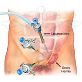
The hernia is protrusion of the body contents through the weakness within the muscle. It is logical that something originating from inside is better dealt from the inside. Also by doing this one does not cut and weaken the already weak muscles at the hernia site. In laparoscopic surgery we use ultra light weight mesh and it is introduced with 10 mm small hole.
Use of mesh in hernia is standard procedure even in open surgery. The mesh used is equivalent to the main one employed for open operations over last 3 decades. Its safety and efficacy is certainly as proved through the numerous trials in the USA and Europe. Q. Is laparoscopic surgery very costly? How can a patient justify the cost of the equipment and laparoscopic surgery? A. The initial cost of setting up of laparoscopic instrument of high definition is about rupees twenty lakhs. Once the initial setting up expenditure of laparoscopic surgery is included, the cost of surgical treatment can be minimized by using re-usable laparoscopic instrument which is now being made in all the developing country.
All the patients who get laparoscopic surgery are benefited. The working person who returns to work quickly has tremendous benefit for that self-employed. A poor labour can resume work or return to home soon and take control from the disrupted hand to mouth as may the situation be. Young children are in a position to return to school soon and do not lose out on studies or sport.
Employers are in great benefit, it means fewer sick leaves and early return back to work e.g. following a gallbladder operation, a worker finds it hard to resume work till about about six weeks to three months. Here, they can be back to work in a week or two.
With the advancement of technology, the engineers and manufacturers have responded with telescopes of smaller diameter like 5 mm and three mm as opposed to the ‘conventional’ laparoscopic 10mm telescopes. Also instruments are now being developed of 3 mm diameter. This advancement is known as mini/micro/needloscopic laparoscopic surgery. The other advancement is Single Incision Laparoscopic Surgery.This is going to be the technique of 21st century. While gallbladder and appendix operations can be achieved with similar equipment hence cost remains the same, advanced laparoscopic surgery like weight reduction bariatric surgery would want special instruments. Robotic surgery, computer-assisted surgery, and robotically-assisted surgery are terms for technological developments that use robotic systems to aid in surgical procedures. Other advancement in laparoscopic surgery is robotically-assisted surgery was developed to overcome both the limitations of minimally invasive surgery or to enhance the capabilities of surgeons performing open surgery. In the case of robotically assisted minimally invasive surgery, instead of directly moving the instruments, the surgeon uses one of two methods to control the instruments; either a direct tele-manipulator or by computer control.
The risk comes from the inexperienced laparoscopic surgeon as there is rarely a more experienced person available for guidance in the event of difficulty. Unlike developed country there is no training program here and all sorts of depends upon individual enterprise. The safer surgeons don’t contemplate it an insult for their ego if they have to transform a laparoscopic procedure to open in the event of difficulty. Aside from this, the only real other thing may be the decrease in sympathy levels from relatives as the hospital stay is so short. Unlike most other professions, changes within the profession of medicine are met with a few resistance and scepticism. Successful examples along with a positive approach are essential for that implementation of such programs. This figure should rise with rise in awareness amongst general practitioners and the public. The near future generations while reading a brief history of surgery will wonder why operations were done open.
Laparoscopic Nissen’s Fundoplication
Laparoscopic Lap Esophagectomy with Gasto Esophagoctomy
Laparoscopic Oesophagus – Heller’s Cardiomyotomy
Stomach
Laparoscopic Gastrectomy
Laparoscopic Gastrojejunostomy
Laparoscopic Peptic Perforation Repair
Laparoscopic Pyloroplasty
Laparoscopic Duodenotomy
Laparoscopic Duodenal Perforation Repair
Laparoscopic Closure Of Bowel Perforation
Laparoscopic Enterotomy Small Bowel
Laparoscopic Excision Of Entero Cutaneous Fistula
Laparoscopic Excision Of Gist Of Small Bowel
Laparoscopic Excision Of Lad’s Band
Laparoscopic Excision Of Meckel’s Diverticulum
Laparoscopic Excision Of Sarcoma Of Small Bowel
Laparoscopic Excision Of Vitello Intestinal Duct
Laparoscopic Jejunostomy
Laparoscopic Abdominal Adhesiolysis
Laparoscopic Small Bowel Resection
Laparoscopic Mesenteric Cyst Excision
Laparoscopic Mesenteric Lymph Nodes Excision Biopsy
Laparoscopic Small Bowel And Mesenteric Pexy
Laparoscopic Small Bowel Resection And Anastomosis
Laparoscopic Small Bowel Strictureplasty
Laparoscopic Anterior Resection
Laparoscopic Hemicolectomy
Laparoscopic Colectomy – Total With Ileo-Rectal Anastomosis
Laparoscopic Hemicolectomy – With Anastomosis
Laparoscopic Hemicolectomy – With Formation Of Stoma
Laparoscopic Proctocolectomy With Ileoanal Pouch And Defunctioning Loop Ileostomy
Laparoscopic Anterior Resection
Laparoscopic Sigmoidcolectomy
Laparoscopic Appendicectomy In Mucocoele
Laparoscopic Drainage Of Appendicular Abcess
Laparoscopic Appendicectomy Surgeon Charges
Laparoscopic Omental Biopsy
Laparoscopic Omentectomy
Laparoscopic Retroperitoneal Abcess Drainage
Laparoscopic Retroperitoneal Cyst Excision
Laparoscopic Retroperitoneal Lipoma Excision
Laparoscopic Retroperitoneal Liposarcoma Excision
Laparoscopic Retroperitoneal Lymph Nodes Excision Biopsy
Laparoscopic Retroperitoneal Scopy
Laparoscopic Peritoneal Metastasis Biopsy
Lap. Prolapse Of Rectum – Abdominal Rectopexy
Laparoscopic High Anterior Resection Of Rectum
Laparoscopic Low Anterior Resection Of Rectum
Laparoscopic Ultra Low Anterior Resection Of Rectum
Prolapse Of Rectum – Laparoscopic Rectopexy
Lap Adrenal Gland Excision
Laparoscopic Splenectomy
Laparoscopic Large Splenectomy
Lap Phrenicoplasty
Laparoscopic Diaphragmatic Hernia
Laparoscopic Epigastric Hernia
Laparoscopic Femoral Hernia
Laparoscopic Incisional Hernia
Laparoscopic Inguinal Hernia
Laparoscopic Paraumbilical Hernia
Laparoscopic Supraumbilical Hernia
Laparoscopic Umbilical Hernia
Laparoscopy is a minimally invasive procedure used to detect and treat abdominal and pelvic disorders. During diagnostic laparoscopy, a long, narrow, flexible tube (laparoscope) is inserted into the abdominal cavity via a small skin incision. A camera is attached to the end of the laparoscope which is inserted inside the body. It captures images of the abdominal organs, and a computer screen connected to the camera displays these images. The surgeon manoeuvres the other end of the laparoscope to detect any pathological changes that have occurred within. Laparoscopy is helpful in diagnosing disorders of all parts of the digestive tract, the liver, the pancreas and pelvic organs as well.
This procedure is done to remove inflamed infected appendix and to prevent perforation of the inflamed appendix into the abdominal cavity and spreading infection. This procedure can be done laparoscopically with small skin incisions which causes less postoperative discomfort and aids in early return to daily activities.
Nissen’s Fundoplication is a surgical procedure performed to treat severe Gastroesophageal Reflux Disease (GERD), known as hyperacidity in layman terms. This surgical procedure has been used successfully to revert the structural anomaly resulting due to a hiatus hernia. During Open Nissen’s Fundoplication, the surgeon makes a single large surgical incision over your abdominal skin. The upper part of the stomach which has herniated above the diaphragm and into the chest cavity is relocated to its former position. The upper curve of the stomach is then wrapped around the lower part of the esophagus tightly enough to allow normal passage of food but to prevent the acid reflux or herniation of the stomach from occurring again.
Splenic abscess forms due to bacterial infection of the spleen or as a result of pre-existing endocarditis. The abscess is typically located with the help of a contrast dye and CT studies. Percutaneous drainage is done in most cases of splenic abscess unless it is contra-indicated. Open surgery may be needed to drain a multi-loculated abscess if there is a risk of haemorrhage.
Splenorrhaphy is a term used for the surgical procedure performed to repair and preserve the structure and function of a damaged spleen. The spleen is a highly vascular organ making it susceptible to considerable damage and bleeding following any trauma.
Splenorrhaphy is preferred over the surgical removal of the entire spleen since it is a very important organ that maintains a person’s immune system. The damaged or torn portion of the spleen is sutured together and repaired to a relative degree of normalcy during the surgery.
Splenectomy is the surgical removal of the entire spleen or a part of it. The spleen may get damaged due to perforating injury or direct trauma with a blunt object. Due to its highly vascular nature, the resultant blood loss and tissue damage could be significant. If splenorrhaphy is contra-indicated, then the surgeon is left with no choice but to remove the entire spleen.
During the surgery, the spleen is surgically separated from the peritoneum, adjacent blood vessels, muscle attachments and removed from the abdominal cavity. The splenic vein and artery are sutured to prevent haemorrhage.
Retroperitoneal cysts develop in the retroperitoneal space and may compress the organs like the kidneys or the spleen if they grow in size. MRI guidance helps to locate the cysts, and they are often drained using minimally invasive techniques. If there is suspicion of a lipoma or a liposarcoma, a preoperative needle biopsy may be performed to confirm the diagnosis.
During the surgery, the retroperitoneal space is explored to determine the area affected by the cysts. The cyst walls are removed along with any remnant debris. Tissue may be obtained for biopsy during the surgery if there is suspicion of malignancy. In the case of lipoma and liposarcoma, the entire structure is resected with precaution to avoid manipulating its integrity. The entire lipoma or liposarcoma may be sent for biopsy.
The Retroperitoneal space is rich in blood and lymph supply, therefore cancer of any of the retroperitoneal organs may spread easily via the space.
If the surgeon suspects such an occurrence then the retroperitoneum is surgically explored to look for signs of malignancy. The lymph nodes are surgically excised, taking care not to damage their structure for the fear of spreading the malignancy. The nodes are then sent for biopsy which helps in detecting and staging cancer.
The retroperitoneal space is rich in blood supply due to the presence of several important blood vessels. Blunt trauma to the retroperitoneal organs may result in haemorrhage and the resultant formation of a haematoma (aggregation of blood outside the blood vessels).
CT helps the surgeon to determine the severity and extent of the haematoma. In case of active bleeding and expanding hematoma surgical exploration is done to stop bleeding.
© 2026 All Rights Reserved